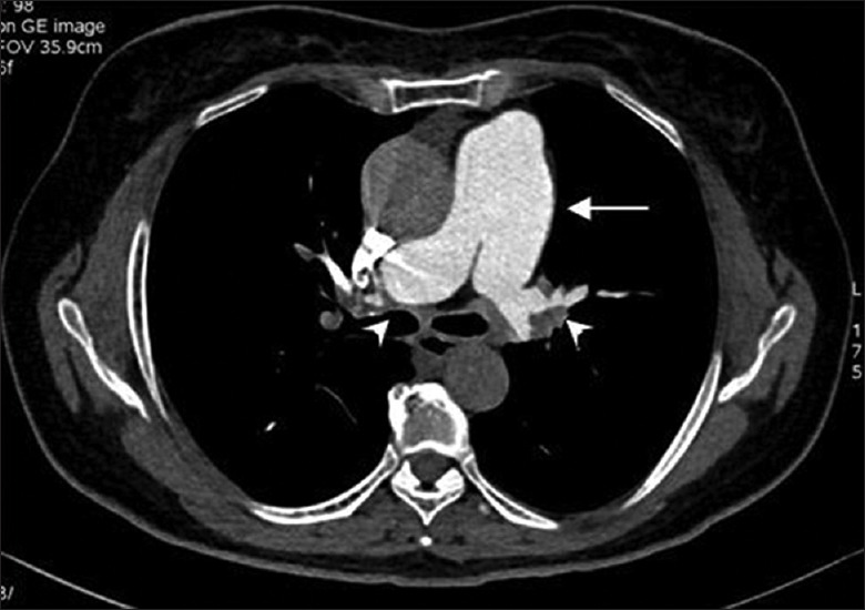
Figure 2.

FM in a 70-year-old woman who presented with dyspnea on exertion and had a positive result of T-SPOT.TB. CT pulmonary angiogram shows widening of the pulmonary artery trunk (arrow) and compression of pulmonary arteries by surrounding soft tissue at the hila (arrowheads). CT: Computed tomography; FM: Fibrosing mediastinitis.
