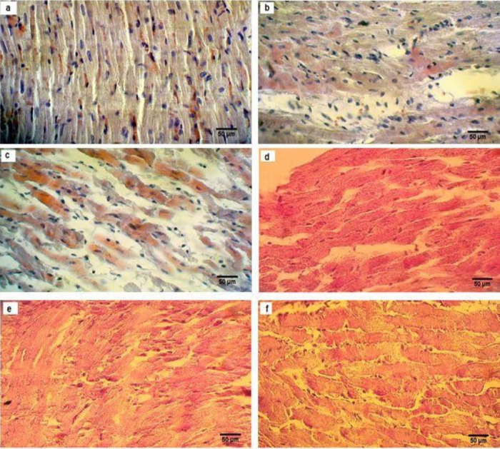
Figure 6.

Histological evaluation of H&E staining (a, b, c) and periodic acid-Schiff (PAS) (d, e, f), examined and analyzed by a light microscope (magnification 40×) in the heart of sham-ovariectomized (OVX) and diabetic ovariectomized (OVX.D) rat groups. Results showed histological changes in sham (a), OVX (b), OVX.D (c), and PAS showed different storage of glycogen in sham (d), OVX (e), and OVX.D (f) animal groups
