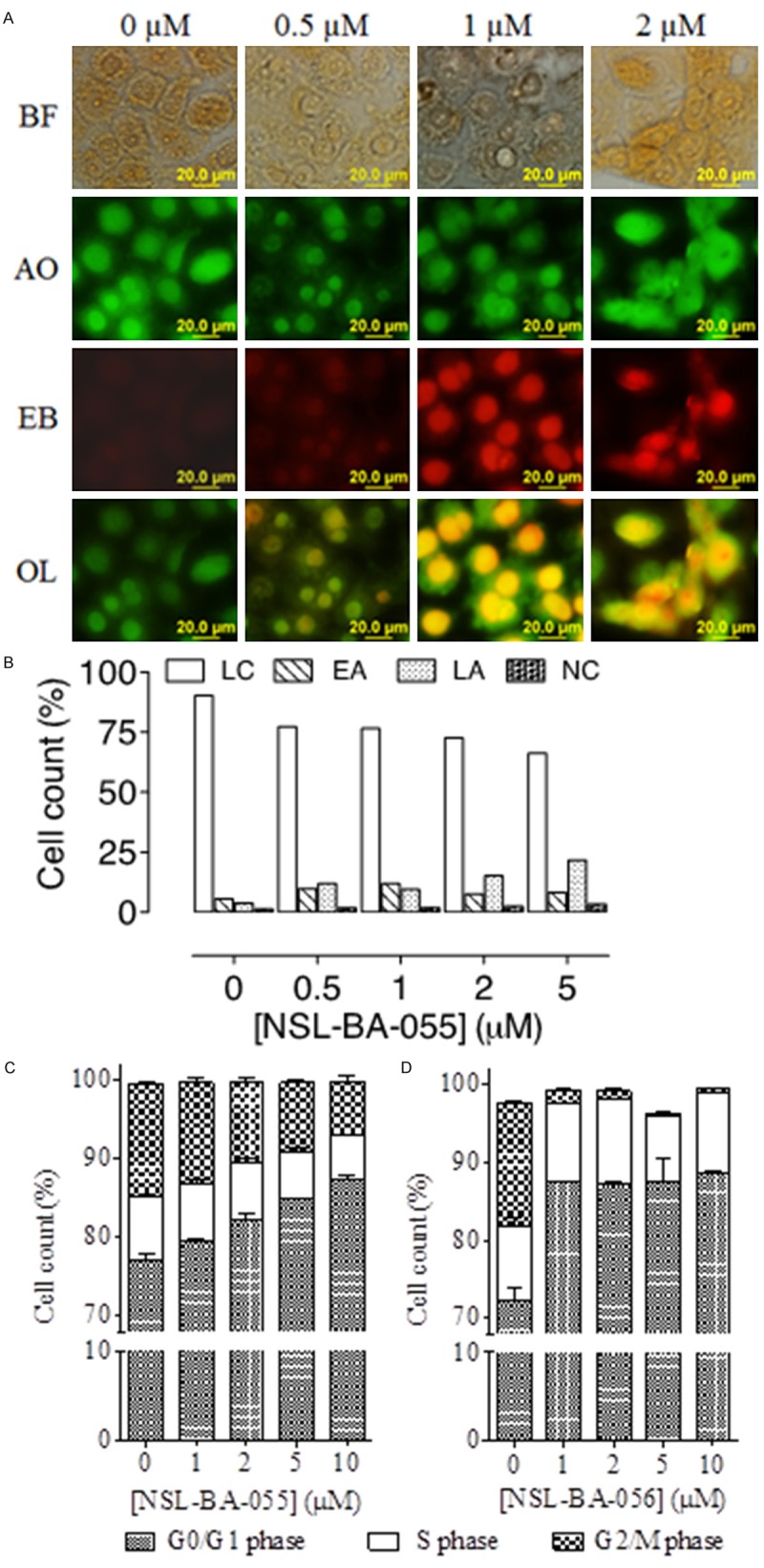
Figure 4.

PCAIs induce apoptosis in pancreatic cancer cells by arresting growth at the G0/G1 phase. A: BxPC-3 cells were treated with NSL-BA-056 for 48 h, stained with 10 µg/mL AO/EB and images captured and analyzed as described in the methods. AO stained live cells green and EB stained dying cells with compromised membranes red. BF, Bright Field; AO, Acridine Orange; EB, Ethidium Bromide; OL, overlay of AO and EB images. B and C: MIAPaCa-2 cells were treated with 0-10 µM of either NSL-BA-055 or NSL-BA-056 for 48 h, stained with Vindelov’s reagent as described in the methods and analyzed using a C6 flow cytometer as described in the methods. The relative percent cell counts in each phase of the cell cycle after treatment was plotted. D: MIAPaCa-2cells were treated with NSL-BA-055 for 48 h, stained with FITC-labeled Annexin V and propidium iodide and analyzed as described in the methods. The results are the means (± SEM, N = 4) and are representative of four independent experiments.
