Figure 5.

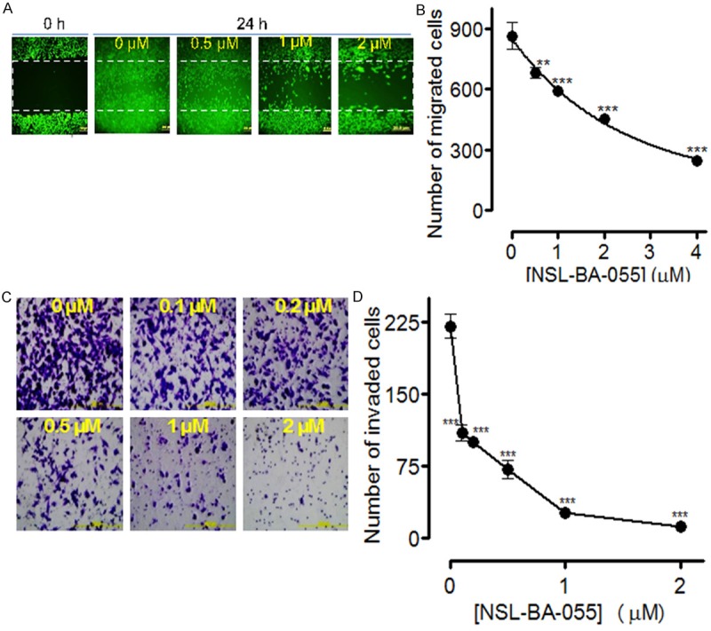
PCAIs inhibit pancreatic cancer cell migration and invasion. A: MIAPaCa-2 cells were incubated in 12-well plates for 24 h, a wound was created with pipette tip, and the cells treated with the indicated concentrations of the NSL-BA-055. After 24 h, the extent of closure was monitored under the microscope and photographed. Representative images of four independent experiments conducted in triplicates are shown. B: MIAPaCa-2 cells were treated with the indicated concentrations of NSL-BA-055. C and D: The cell invasion assay was performed in the Matrigel invasion chambers as described in the methods. All Representative images were 10 × magnification and all data of invaded cells were represented as ± SEM, N = 8-10. Statistical differences between control and treated groups were determined by Dunnett’s post-test comparisons. Significance was defined as *P < 0.05; **P < 0.01 and ***P < 0.001.
