FIG 6.
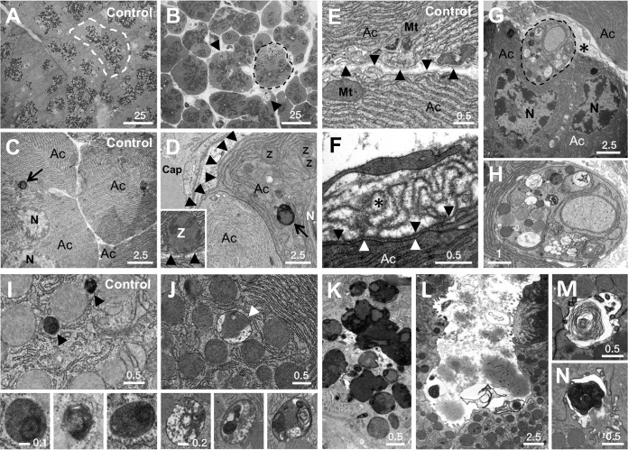
Ultrastructural changes in PTF1A-deficient pancreas. All panels are of 14-day cKO mice except for TAM-treated Ptf1a+/CreER controls in panels A, C, E, and I. (A to D) Effects on apical-basal polarity; dashed lines outline individual acini. Zymogen granules (dark dots), normally clustered apically around lumina (A), are decentralized in cKO pancreas (B; arrowheads indicate phagosomes). Zymogen granules, rarely basal to nuclei (C), are basal in cKO pancreas (D). The inset shows zymogen granule apposed to basal plasmalemma. Arrowheads, basal lamina; arrows, lysosomes. (E and F) Excessive basal lamina (asterisk) of a shrunken acinar cell (F) compared to normal acinar cells (E). Black arrowheads indicate basal lamina from which folds emanate; white arrowheads indicate basal plasmalemma. (G) Phagocytic engulfment by acinar cells (dashed outline in panels B and G). (H) Thin band of cytoplasm bounded by plasmalemma is enlarged. (I) Zymogen granules and lysosomes (arrowheads) in control cells. Insets show enlarged lysosome images. (J) Crinophagic vesicles (arrowhead) are the size of zymogen granules. Insets show complex interior structures. (K) Extensive crinophagic fusions. (L to N) Frequent luminal debris, including lamellar membranes. Size bar length units are micrometers. Ac, acinar cell; Cap, capillary; Mt, mitochondrion; N, nucleus; Z, zymogen granule.

