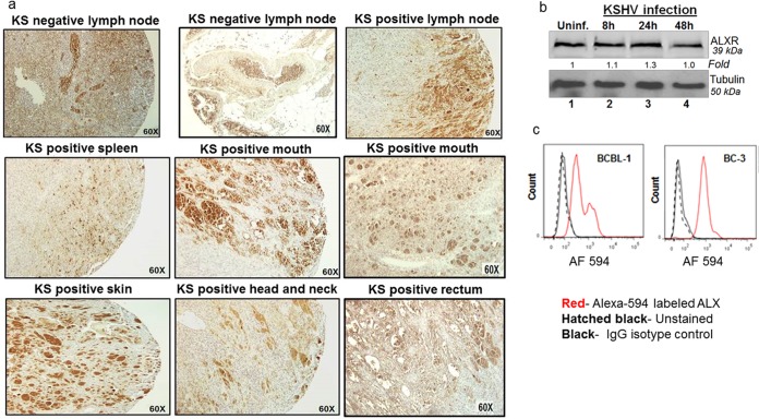
FIG 2.
Expression of lipoxin receptor ALX/FPR on human KS tissue sections and in vitro cell models of KS and PEL. (a) A KS tissue section array from the ACSR was immunohistochemically stained for ALX/FPR, and microscopy was performed. The brown color indicates ALX/FPR staining. (b) Lysates from HMVEC-d KSHV-infected cells (30 DNA copies/cell) for the indicated time points (8, 24, and 48 h) and uninfected control HMVEC-d cells were Western blotted for ALX/FPR, stripped, and immunoblotted for tubulin. (c) In vitro PEL models BCBL-1 and BC-3 cells were fixed and stained for ALX/FPR using an Alexa 594-labeled antibody and, as a control, stained for IgG isotype.

