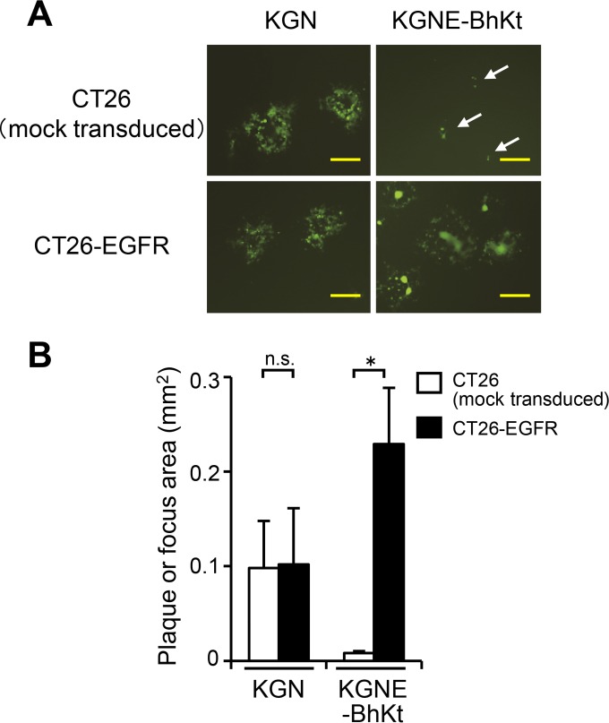
FIG 6.
Specificity of lateral spread by KGNE-BhKt. (A) CT26-EGFR cells were infected with the viruses indicated above the panels (MOI of 10). Extracellular viruses were inactivated by an acidic wash, and equal numbers of infected (donor) cells were added to monolayers of the uninfected (acceptor) cells indicated to the left. The mixed cultures were overlaid with methylcellulose-containing medium, and EGFP signals were recorded at 2 days postinfection. Bars, 500 μm. Arrows show single green cells or small foci. (B) Mean areas of plaques or foci (n = 15) in the wells examined for panel A. Error bars represent standard deviations. White bars, CT26 (mock-transduced) cells; black bars, CT26-EGFR cells. *, P < 0.05 by the Welch t test; n.s., not significant.

