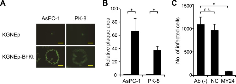
FIG 7.
Lateral spread of syncytial, EpCAM-retargeted HSV on human cancer cells. (A) The cell lines listed above the panels were infected for 2 h with the viruses indicated to the left and then overlaid with methylcellulose-containing medium. EGFP signals were recorded at 3 days postinfection. Photographs of representative plaques are shown. Bars, 500 μm. (B) Mean areas of the plaques (n = 15) from panel A normalized to the respective means of KGNE plaque areas. Error bars represent standard deviations. White bars, KGNEp; black bars, KGNEp-BhKt. *, P < 0.05 by the Welch t test. (C) Inhibition of KGNEp-BhKt entry by pretreatment of AsPC-1 cells with 100 μg/ml anti-EpCAM MAb MY24 or an isotype-matched negative-control antibody, as indicated below the columns. Pretreated cells were incubated with KGNEp-BhKt at an MOI of 0.01 for 2 h, extracellular viruses were inactivated, and cells expressing EGFP were counted at 12 h postinfection. Means for 3 replicates are shown, and error bars represent standard deviations. Ab (−), no antibody; NC, isotype-matched negative-control antibody (MG1-45). *, P < 0.05 by the Welch t test; n.s., not significant.

