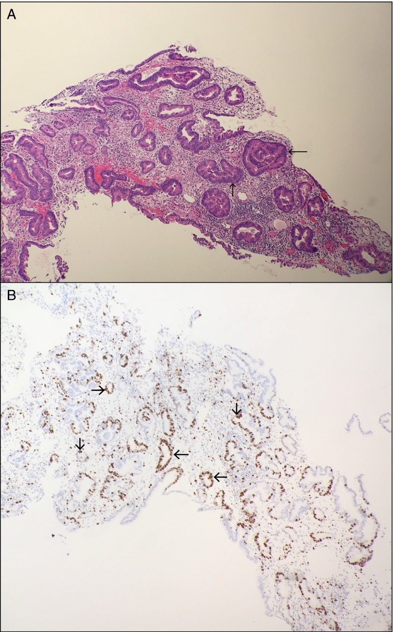
Figure 2.
(A) Hematoxylin and eosin staining of the specimen revealed adenomatous glands with high-grade dysplasia, as manifested by nuclear atypia (arrows). (B) Proliferation marker MIB1 showed strong nuclear staining in dysplastic cells (arrows). There is no evidence of dysplastic cells beyond the basement membrane.

