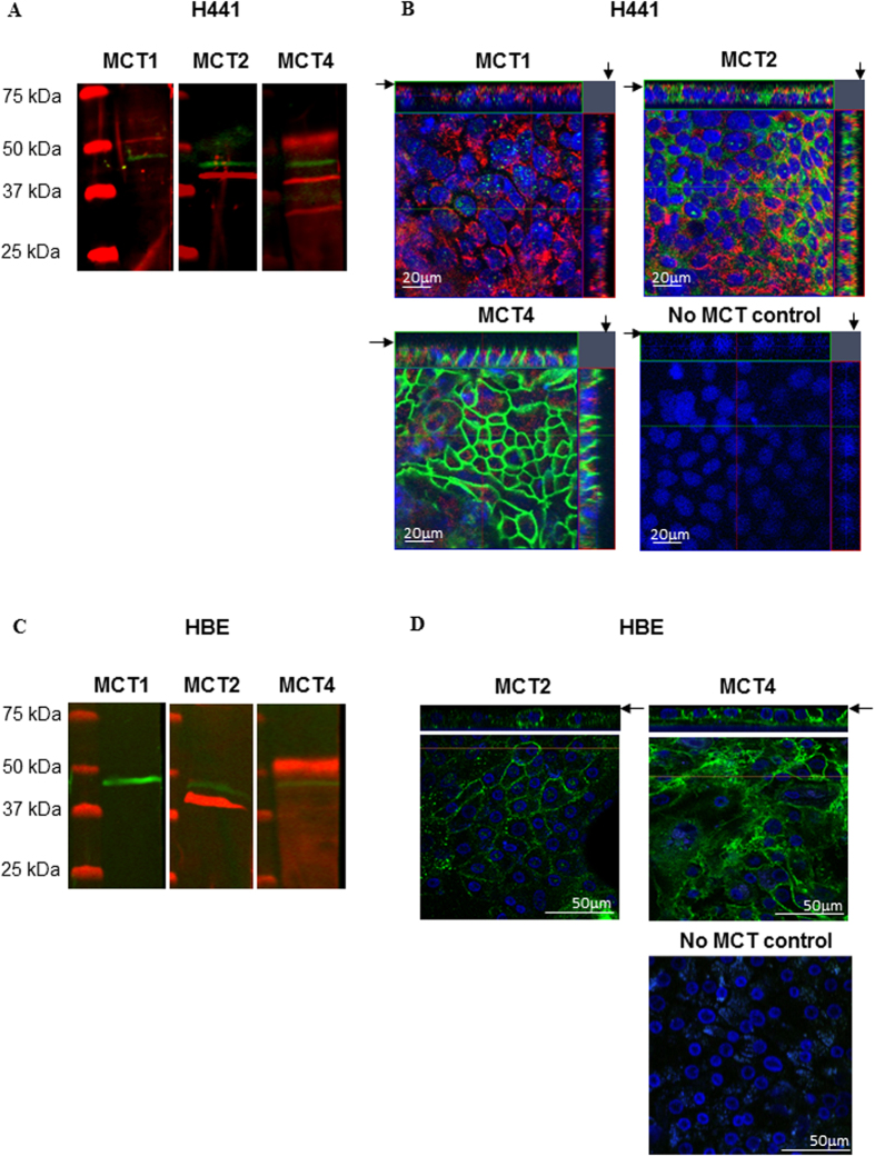
Figure 4. Airway epithelia express monocarboxylate transporters (MCT) 2 & 4.
Western blot and confocal images of immunostained MCT in H441 (A,B) and HBE (C,D) cells grown on Transwell supports. (A,C) Western blots of MCT1 (not detected in HBE), 2 and 4 (red) and β-actin (green). (B,D) XY planes (centre) and XZ planes to the top and right (H441 only); apical surface indicated by an arrow; biotin-conjugated fluorophore labelling of the apical surface (red) (H441 only), MCT1, 2 and 4 (green) or absence of primary antiserum (no MCT control), DAPI stained nuclei (blue).

