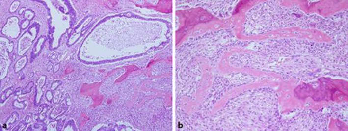
Fig. 3.
Histological analysis of the excised tumor. a Moderately differentiated adenocarcinoma with numerous foci of osteoid and ossification (hematoxylin and eosin stain. ×40). b Osteoblasts are linked by tight and gap junctions and integrated with the underlying osteocyte, namely osteoblastic rimming (hematoxylin and eosin stain. ×100).

