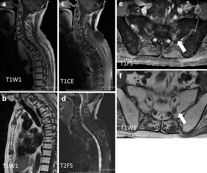
Fig. 1.
Sagittal T1-weighted MRI of the cervical and thoracic spine (a) and sternum (white circle) (b) showed several hypointense lesions. Sagittal T1-weighted contrast-enhanced MRI (c) showed that low-intensity lesions of the vertebrae lacked contrast effects. On sagittal fat-saturated T2-weighted images (d), some lesions were not seen because their signal was equivalent to that of adjacent marrow. The sacral lesion, which was detected by positron emission tomography/computed tomography, had a contrast effect (white arrow) on T1-weighted contrast-enhanced MRI (e) but was not seen on fat-saturated T1-weighted images (f).

