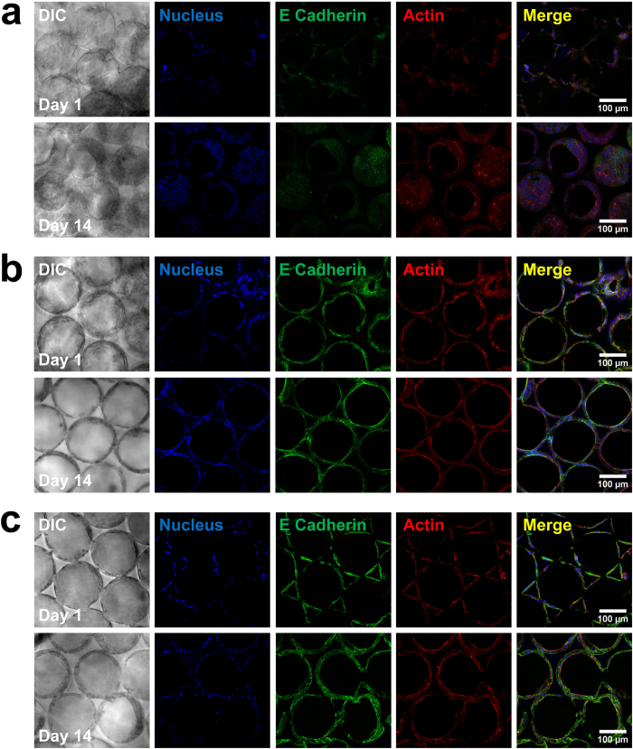
Figure 6.
Immunofluorescence staining of E-cadherin in Huh-7.5 cells on (a) the bare PEG-DA scaffold, (b) the collagen-functionalized scaffold, and (c) the fibronectin-functionalized scaffold, on days 1, 7, and 14. E-cadherin was stained with Alexa Fluor 488 (green), F-actin was stained with Alexa Fluor 555 phalloidin (red), and cell nuclei were counterstained with DAPI (blue). High quality figures of the day 14 merged images are shown in Supplementary Fig. S2.

