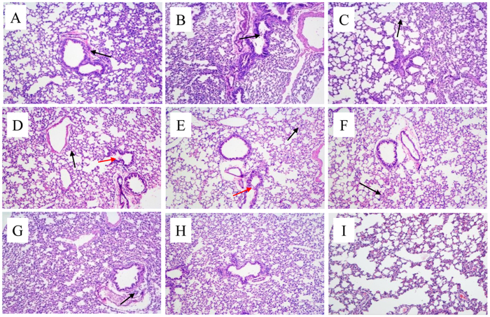
Figure 7. Microscopic lung sections from mice inoculated with H4 viruses at 5 dpi.
(A–D): 29-1/H4N2, MH-2/H4N6, 44-2/H4N6, 46-2/H4N6; E-H): 67-2/H4N6, 408-1/H4N6, 420-2/H4N6, 421-2/H4N6; (I): Mock. Lungs were harvested at 5 dpi from mice inoculated intranasally with 106 EID50 viruses. The black arrow represents infiltration of inflammatory cells, and the red arrow indicates mild detachment of bronchial epithelial cells.

