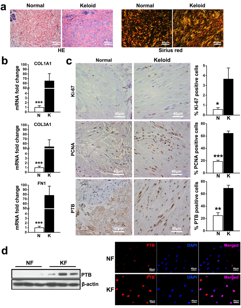
Figure 1. Accumulated ECM, increased cell proliferation and upregulated PTB expression were detected in keloid.
(a) HE staining and sirius red staining were conducted for keloid tissues and normal skin. (b) The expression of COL1A1, COL3A1 and FN1 in dermal tissues was evaluated by real-time PCR. Data are shown as the mean ± SEM, n = 15. ***P < 0.001 by student’s t-test. (c) The expression of Ki-67, proliferating cell nuclear antigen (PCNA) and PTB was tested using immunohistochemistry analysis. Data are shown as the mean ± SD, n = 15. *P < 0.05, **P < 0.01, ***P < 0.001 by student’s t-test. (d) Passage three of fibroblasts were harvested from normal skin and keloid tissues to detect the expression of PTB by western blot and immunofluorescence, respectively. In western blot analysis the expression of β-actin was used as an internal control for protein loading normalization. N: normal skin; K: keloid tissue. NF: fibroblasts from normal skin; and KF: fibroblasts from keloid tissues.

