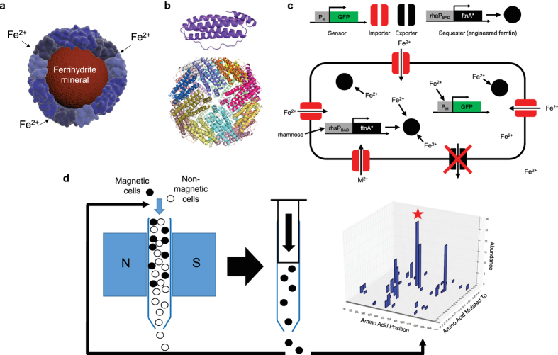
Figure 1. Engineering cellular magnetism.
(a) Schematic of the ferritin protein shell encaging a ferrihydrite nanoparticle (b) crystal structure of the ferritin monomer (top) and the self-assembled, 24-homomers cage (bottom). (c) The cell engineered to accumulate metals by knockout of genomic exporters (black) and expression of importers (red). Mutant ferritins particles (black spheres) induced by a rhamnose promoter biomineralize iron into intracellular magnetic particles. A genetic fluorescence sensor monitors intracellular free Fe2+ level. (d) directed evolution for increased magnetism: iterative selection by high gradient magnetic column of a library of cells expressing randomly mutated ferritins was carried out over 10 days (1 cycle/day). Subsequent sequencing analysis of the magnetically retained mutants enabled discovery of mutations in key residues (e.g. red star: T64) that enhanced cellular magnetism.

