
Figure 3. Reduced density of PNNs in peri-infarct areas.
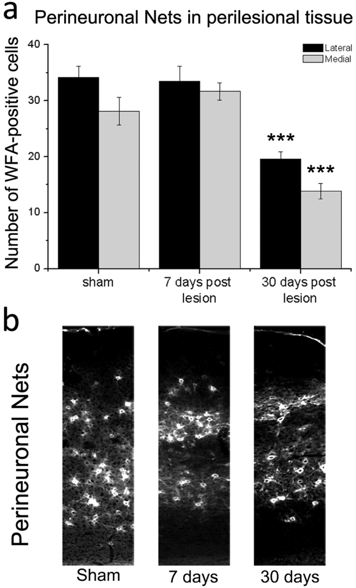
(a) Number of cells surrounded by PNNs in 200 μm wide cortical columns, lateral (black bars) and medial (grey bars) to the ischemic lesion, 7 (n = 5) and 30 days post injury (n = 8). Similar cortical regions were also sampled in controls (n = 8). PNN density is significantly reduced at 30 but not 7 days after stroke (one Way ANOVA, post hoc Holm-Sidak test, ***p < 0.001). Data are mean ± SE. (b) Representative images acquired from coronal sections of the motor cortex from sham and stroke mice at 7 and 30 days. Column width = 200 μm.