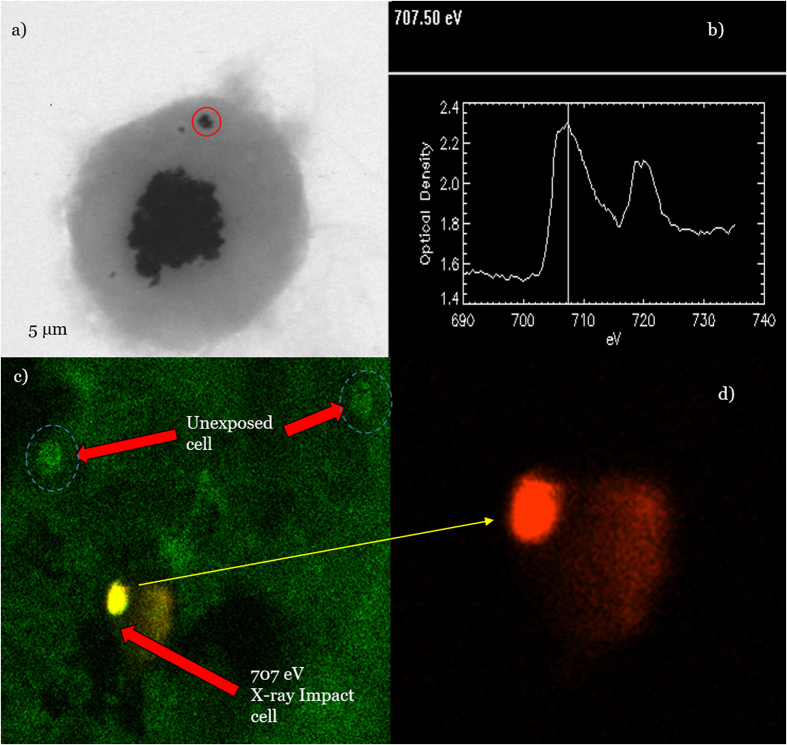
Figure 7.
STXM image of a macrophage containing nanoparticles (red circle) selected for X-ray irradiation (a) and the corresponding 705 eV X-ray absorption spectrum (b) of the selected nanoparticle site (red circle). Oxidant-DHE fluorescence image of a macrophage containing IONs, which was irradiated using a 30 nm-focused X-ray photon of 707 eV. Photoelectric-absorption LEE emission demonstrated energy transfer to local oxygen, producing a distribution of  via unfiltered (c) and filtered red fluorescence (d) of the oxidant DHE in unexposed and exposed cells and in an exposed cell alone, respectively. The material was mostly confined within a single cell, but trace secondary radiolysis of water by fluorescent X-rays was feasible.

