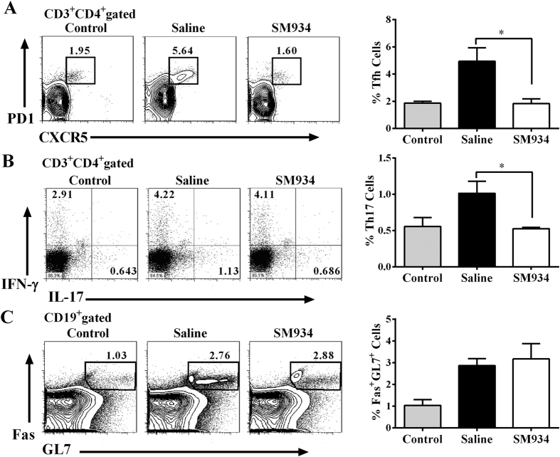
Figure 4. SM934 restrained Tfh and Th17 cells development but not GC B cells in CIA mice.
Flow cytometric analysis of Tfh cells by PD-1 and CXCR5 staining (A), Th17 and Th1 cells by IL-17 and IFN-γ staining (B), or GC B cells by Fas and GL7 staining (C) in splenocytes from CIA mice. The representative (left) and statistical results (right) were showed. Values are the mean ± SEM. *p < 0.05 compared with the saline-treated CIA mice (saline). Results are derived from 2 independent experiments with similar pattern in each treated group (n = 8 in each group).

