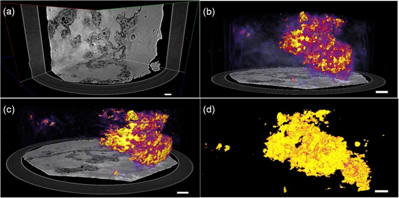
Figure 3. Three-dimensional renderings of PR-based SR-PPCT scans of different human HAE lesion components.
(a) Virtual cut through the central volume of the HAE specimen in Fig. 4(d), showing the different HAE pathological feature structures and connectivity. (b) Delineation of the volumetric distribution of the HAE lesion, parenchyma and halo zone (marginal zone), surrounding the lesions, (c) shows the 3D morphology of fibrosis and calcification of lesion tissues, and (d) shows the volumetric distribution and 3D shape of the completely calcified lesions. The length of the scale bar is 150 μm.

