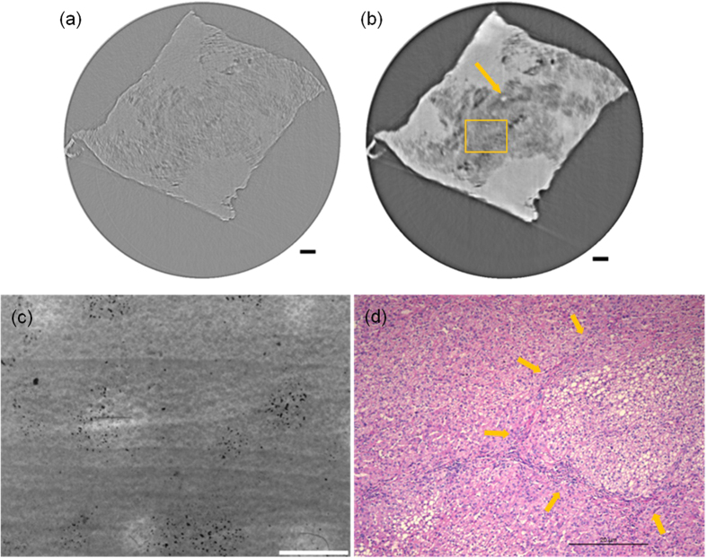
Figure 4.
Comparison and evaluation of higher resolution SR-PPCT sectional image with a slice thickness of 1.625 μm and its histopathologic examination image of a local HAE lesion, corresponding to the rectangle region 3 in Fig. 1(a,d). Reconstructed sectional images using the SR-PPCT technique (a) and PR-based SR-PPCT technique (b,c) is the enlarged view of the rectangle plotted in (b,d) the H&E stained image under a light microscope (400×). The length of the scale bars are 80 μm in (a,b), and 20 μm in (c,d).

