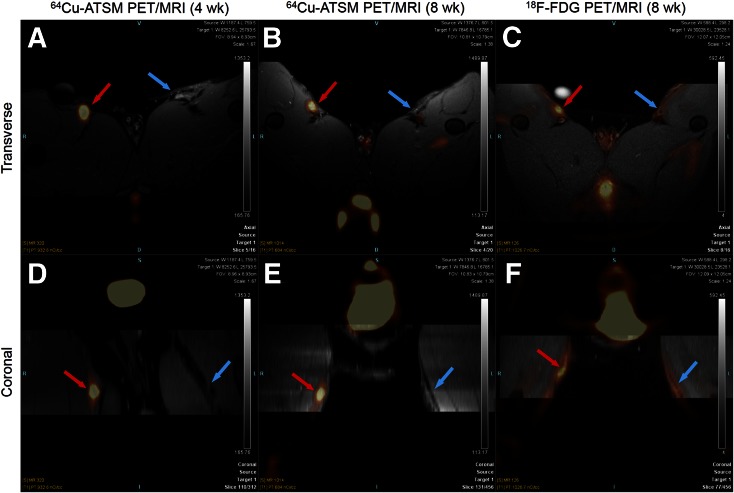
FIGURE 2.
Transverse (top) and coronal (bottom) view of 64Cu-ATSM PET/T1-weighted MR images of representative rabbit at 4 wk (A, D) and 8 wk (B, E) after injury and 18F-FDG PET/T1-weighted MR images of same rabbit 8 wk after injury (C, F). Red arrows point to injured femoral artery; blue arrows point to sham-operated femoral artery.

