FIGURE 4.
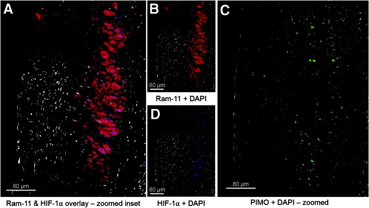
In atherosclerotic plaque of injured femoral artery, HIF-1α staining is localized within areas of high RAM-11–positive macrophages (A), confirmed by viewing single-channel staining of RAM-11 (B) and HIF-1α (D). Detection of pimonidazole (PIMO) adducts was done on adjacent slide (C), and pimonidazole positivity was observed in area of RAM-11+HIF-1α displayed in A, B, and D. A and C are at the same scale. For all panels, the color code is as follows: DAPI, white; RAM-11, red; HIF-1α, blue; PIMO, green.

