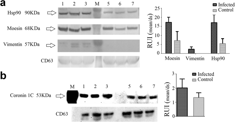
Figure 3. Western blot confirmed proteins significantly more abundant in exosomes from Mtb-infected cells originally detected by LC-MS/MS.
Hsp90, Moesin and Vimentin were detected using a chromogenic substrate (a). The intensity of the bands was evaluated relative to the intensity of CD63. Additionally, Coronin 1 C was detected using a chemiluminescent substrate (b). The intensity of the bands was evaluated relative to the intensity of CD63. Samples from three independent experiments were evaluated. Exosomes from infected cells, lanes: 1, 2 and 3. M: Molecular weight marker. Exosomes from control cells lanes: 5, 6 and 7. The original images of the western blots can be found in Supplementary Fig. S3. RUI: relative units of intensity.

