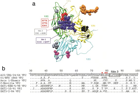
Fig. 2.

Antigenic sites identified in the capsid crystal structure of the FMDV/O1 BFS 1860. a The O1 BFS 1860 asymmetric unit (PDB # 1FOD) was manipulated with Chimera, consisting of 1 VP1 (yellow), 1 VP2 (green), 1 VP3 (cyan) and 1 VP4 (tan), with previously identified antigenic sites shown as blue spheres (site 1), orange spheres (site 2), gray spheres (site 3), black spheres (site 4) and magenta spheres (site 5), and with A22 Iraq escape mutant mutations (H77R, Q79K and K80T) depicted as red spheres, respectively. b Partial amino acid alignment of the VP2 sequences of representative FMDV serotypes. Positions of the FMDV/A22 Iraq escape mutant mutations are indicated as red dots and the adjacent FMDV/A antigenic site 3 is shown in a box
