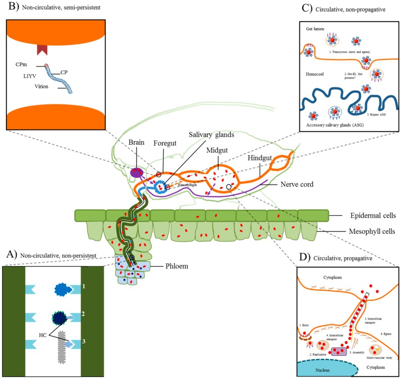
Figure 1.
Plant virus transmission strategies in insect vectors. A viruliferous insect is shown feeding on infected phloem tissue (stylet in dark green). (A) Non-circulative, non-persistent viruses are retained in the distal tip of the insect stylet (small blue hexagons) through two strategies, capsid-only or helper-dependent (Inset A: Magnification of the insect stylet duct region). In capsid-only, virion attachment to the insect stylet is facilitated by a direct interaction mediated by the capsid protein (1). In helper-dependent, attachment of the virion to the stylet is accomplished using several (2; caulimoviruses) or a single non-structural protein(s) (3; potyviruses); (B) Non-circulative, semi-persistent viruses are retained within the insect foregut (Inset B: Magnification of insect foregut). Crinivirus (lettuce infectious yellows virus; LIYV) attachment to the insect foregut appears to be dependent on only the minor capsid protein (CPm; small brown circles); (C) Circulative, non-persistent viruses are non-replicating and require invasion of multiple insect organs to reach the salivary glands for transmission (Inset C: Magnification of luteovirus transmission route from the midgut to the accessory salivary glands). Following ingestion, virions are transported along the alimentary canal and transcytose across the gut (hindgut or midgut) to the hemocoel mediated by the major capsid protein (small red hexagon). However, interaction with and passing through the accessory salivary glands is thought to involve the minor read-through capsid protein (blue loops extending from small red hexagon). Bacterial endosymbionts in the hemocoel have been hypothesized to aid the transmission process for some luteoviruses and geminiviruses by secreting protective proteins; (D) Circulative, propagative viruses replicate and systemically invade several insect organs and tissues with the primary goal of entering the hemolymph or neuronal tissues in order to reach the salivary glands for transmission (Inset D: Magnification of the infection route of rice dwarf virus; RDV). RDV (small red hexagons) enters the midgut by endocytosis in a P2- (outer capsid protein) dependent manner. This is followed by P2-mediated release of RDV virions from endosomes to initiate viral replication and assembly. Virions are then transported cell-to-cell through viral Pns10-derived tubular structures, which bypasses the hemocoel. Modified from [1,4,5,6]. HC, helper component; CP, coat protein; ASG, accessory salivary glands.

