Figure 5.
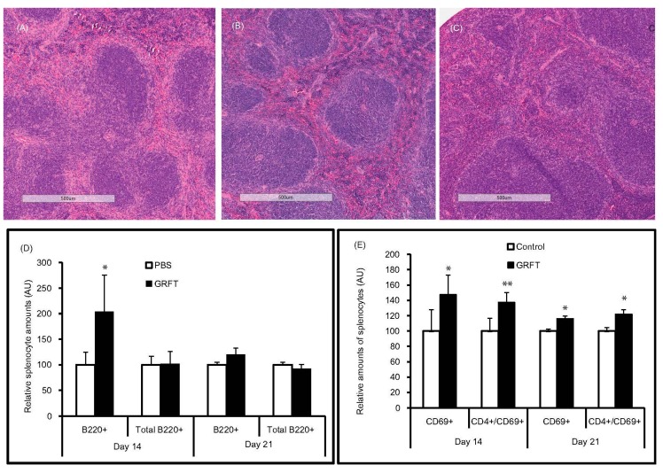
Effect of chronic GRFT treatment on spleen tissue and cell activation. (A–C) hematoxylin and eosin (H and E) stained spleens showing no, to minor, changes in spleen histology after 14 daily treatments with GRFT at 10 mg/kg. (A,B) are representative spleen specimens from the GRFT group at treatment end (Day 14) and Day 21 (14 days of treatment followed by a week of recovery), respectively; (C) representative spleen specimen from the PBS group at Day 14; flow cytometry was carried out after dual fluorescent staining with FITC-conjugated anti-CD4 or CD19 mAb in combination with PE-conjugated anti-B220 (D) or anti-CD69 (E). Relative amounts of splenocytes expressing a given surface marker were obtained with the PBS group value set at 100. B220+, cells staining positive for B220 only (single positive); total B220+ represent B220 single positive and B220/CD19 double positive (*, p < 0.05; **, p < 0.01)) .

