
Fig. 3.
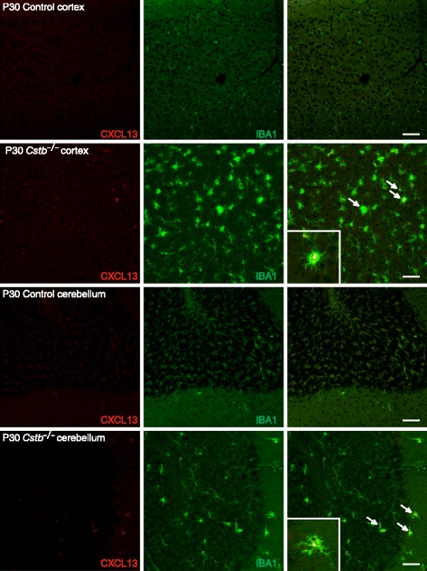
Immunohistochemical detection of CXCL13 in control and Cstb −/− mouse brain at P30. CXCL13-positive microglia are shown by double immunofluorescence staining of CXCL13 (red) with the microglial marker IBA1 (green) in the cortex and cerebellum of control and Cstb −/− mice. Representative CXCL13- and IBA1-double-positive cells in the merged image are marked with arrows. The inserts show enlargements of one double immuno-positive cell from both brain regions. Scale bar = 50 μM