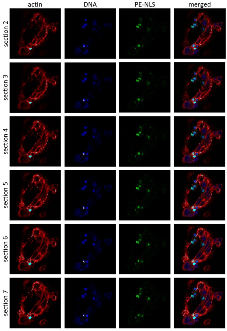
Figure 4.
Localization of PE-NLS in HepG2 cells. Cells, intoxicated with fluorescently labeled PE-NLS (green), were fixed 3 h after treatment and monitored using confocal microscope. The nuclei were labeled with NucRed Live 647 ReadyProbes Reagent (blue), actin was labeled with Alexa Fluor 594 Phalloidin (red). The cells were visualized by confocal microscopy at 60× magnification of the objective.

