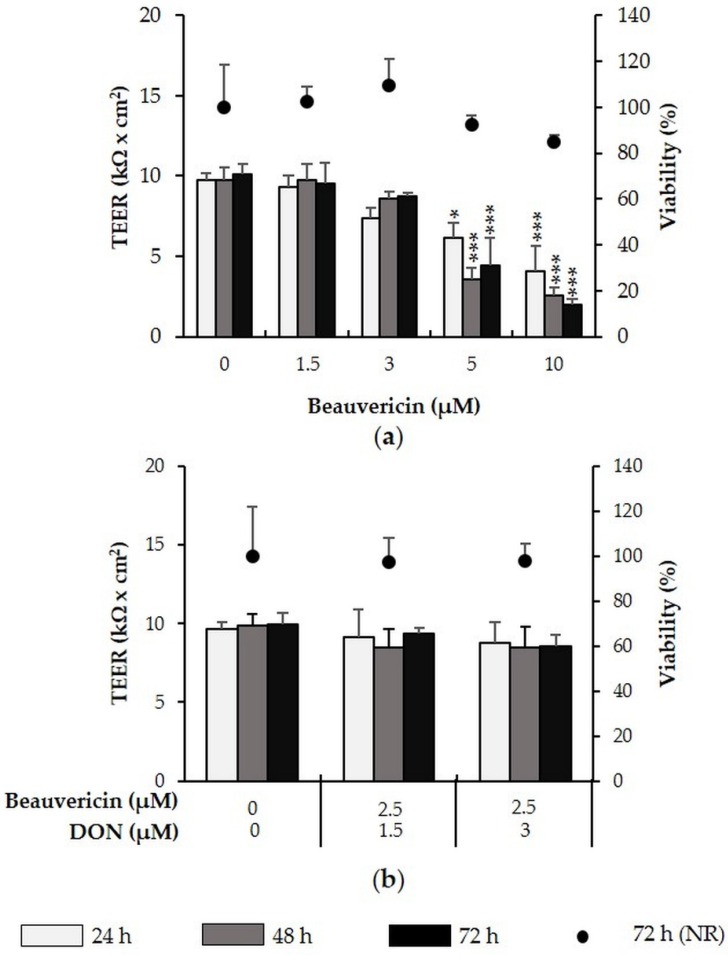
Figure 2.
Effect of beauvericin (+/− DON) on TEER and viability of differentiated intestinal porcine epithelial cells (IPEC)-J2. IPEC-J2 were treated with (a) beauvericin (1.5–10 µM) as well as (b) a combination of beauvericin (2.5 µM) and DON (1.5 or 3 µM). TEER was measured after 24, 48 and 72 h. After the final TEER measurement, viability was determined via the NR assay. Asterisks indicate significant differences compared to control of the respective time point (* p < 0.05, ** p < 0.01, *** p < 0.001). Data represent mean ± SD, n = 3.

