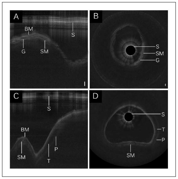
Figure 2.
Long-range optical coherence tomography images of the (A–B) subglottis and (C–D) trachea. Select segments of raw LR-OCT frames of the (A) subglottis and (C) trachea are displayed in Cartesian coordinates. Data conversion to polar coordinates depicts (B, D) the airway in an axial representation. Noise bands were removed for image clarity. Bar = 500 μm.
Abbreviations: BM, basement membrane; G, submucosal gland; P, perichondrium; S, sheath; SM, submucosa; T, tracheal cartilage.

