Fig. S3.
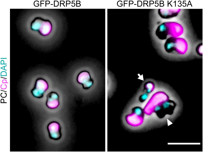
DAPI-stained images of GFP-DRP5B and GFP-DRP5B K135A cells 24 h after the onset of the heat-shock treatment. The arrowhead indicates a cell with one chloroplast and two nuclei. The arrow indicates a cell with the tiny chloroplast remnant. Magenta, autofluorescence of the chloroplast; cyan, DNA stained with DAPI. Images obtained by fluorescence and phase-contrast microscopy are overlaid. (Scale bar: 5 µm.)

