Fig. 1.
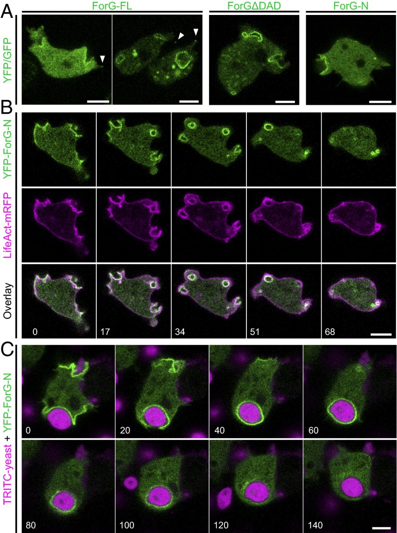
ForG localizes to endocytic structures. (A) GFP-tagged ForG-FL, constitutively active ForGΔDAD (amino acids 1–1,040), and YFP–ForG-N (amino acids 1–423) ectopically expressed in vegetative Dictyostelium cells prominently localized to nascent macropinosomes. Faint enrichment at filopodia tips is indicated by white arrowheads. (B) Time-lapse imaging of cells coexpressing LifeAct-mRFP and YFP–ForG-N revealed that ForG localization is fairly distinct from cortical F-actin. (Bottom) Merged images illustrate striking colocalization of F-actin and ForG at macropinosomes. (C) YFP–ForG-N also accumulated at phagocytic cups throughout engulfment of the large TRITC-labeled yeast particles. Confocal sections are shown in B and C, and correspond to Movies S1 and S2. Time is given in seconds. (Scale bars: 5 μm.)

