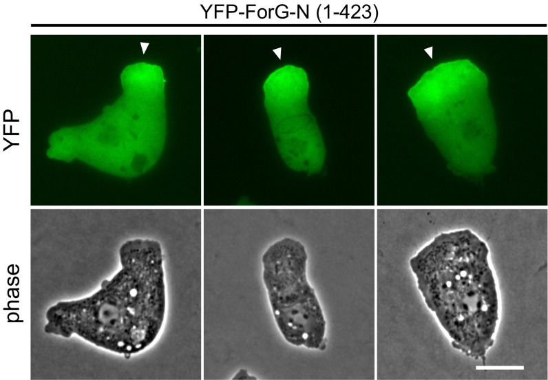
Fig. S1.
ForG accumulates at the leading edge in 2D confinement. To visualize ForG localization in the absence of endocytic cups, the formation of these structures was physically suppressed by overlaying the cells with a thin sheet of agar. In the agar overlay, YFP-tagged ForG-N expressed in WT growth-phase cells accumulated at the leading edge. (Scale bar: 10 μm.)

