Fig. 1.
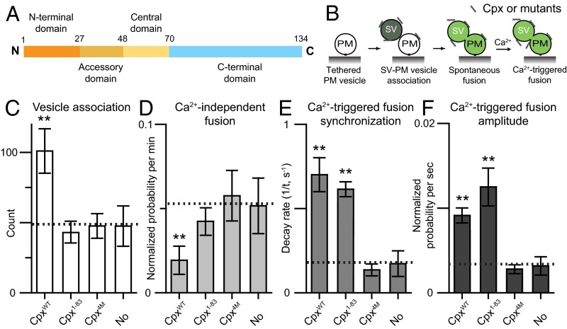
The C-terminal domain of Cpx is important for suppressing Ca2+-independent fusion, but not for Ca2+-triggered fusion. (A) Domain structure of complexin-1. (B) Schematic diagram of the single-vesicle content mixing assay (Methods). The bar graphs show the effects of 2 μM Cpx or Cpx fragments on the SV–PM vesicle association count during the first acquisition periods (Methods) (C), the average probability of Ca2+-independent fusion events per second (D), the decay rate (1/τ) of the histogram upon Ca2+ injection (E), and the amplitude of the first 1-s time bin (probability of a fusion event in that bin) upon 500 μM Ca2+ injection (F). The fusion probabilities and amplitudes were normalized with respect to the corresponding number of analyzed SV–PM vesicle pairs (Methods). Individual histograms are shown in Fig. S1. The dashed lines indicate the control without Cpx. C, D, and F show means ± SD for multiple independent repeat experiments (Table S1). E shows decay rates and error estimates computed from the covariance matrix upon fitting the corresponding histograms with a single exponential decay function using the Levenberg–Marquardt algorithm. **P < 0.01 using the Student’s t test with respect to the control experiment without Cpx (dashed line).

