Fig. 2.
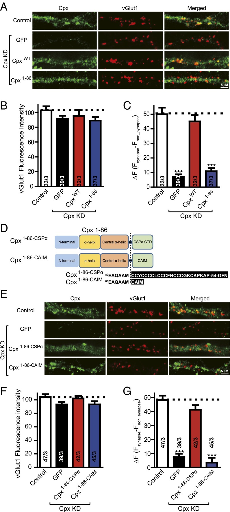
The C-terminal domain of Cpx is important for subcellular location. (A) Representative images of cultured mouse cortical neurons infected with control lentivirus or with lentivirus expressing the Cpx shRNA plus either EGFP only (Cpx KD), or together with wild-type Cpx (CpxWT), or C-terminal truncated mutant Cpx (Cpx1–86). Neurons were fixed and labeled by double immunofluorescence using vGlut1 (to mark excitatory synapses) and Cpx antibodies along with fluorescently labeled secondary antibodies (Methods). The scale bar in right lower corner applies to all images. (B) Summary graphs of vGlut1-specific fluorescence intensities for all conditions as described for A. (C) Summary graphs of ΔF (Fsynapse − Fnon_synapse) of Cpx-specific fluorescence intensity for all conditions as described for A. (D) Domain structures and Cpx chimeras in which the C-terminal sequence was replaced with either the C-terminal sequence of CSPα (Cpx1–86–CSPα) or with the CAIM sequence (Cpx1–86–CAIM). (E) Representative images of cultured mouse cortical neurons infected with control lentivirus or with lentivirus expressing the Cpx shRNA plus either EGFP only (Cpx KD), or together with Cpx synaptic vesicle target mutant (Cpx1–86–CSPα), or Cpx plasma membrane target mutant (Cpx1–86–CAIM). Neurons were treated as described for A. The scale bar in right lower corner applies to all images. (F) Summary graphs of vGlut1-specific fluorescence intensities for all conditions as described for E. (G) Summary graphs of ΔF (Fsynapse − Fnon_synapse) of Cpx-specific fluorescence intensity for all conditions as described for E. Data shown in summary graphs are means ± SEM; numbers of cells/independent cultures analyzed are listed in the bars. Statistical assessments were performed by the Student’s t test comparing each condition to the indicated control experiment (***P < 0.001).

