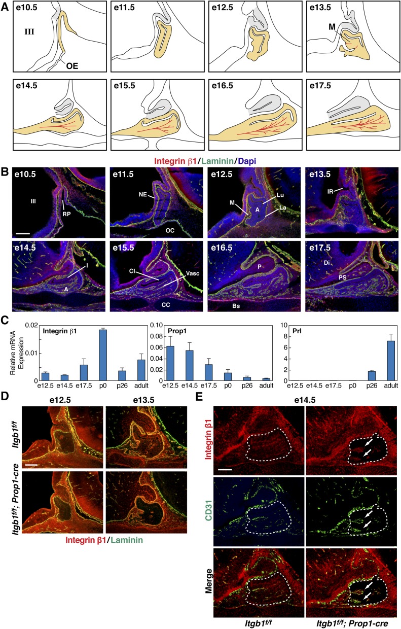
Fig. S1.
Integrin β1 is expressed in pituitary gland epithelial cells throughout embryonic development and is inactivated at e10.5 with Pitx1-cre and at e14.5 with Prop1-cre. (A) Embryonic development of the glandular anterior and intermediate lobes (yellow) beginning with invagination of the oral ectoderm at e10.5. Tandem outgrowth of the third ventricle gives rise to the neural posterior lobe (gray). At e13.5, the first evidence appears of nascent microvessels (M) in red. (B) Immunohistochemical staining of integrin β1 and laminin in sections of control embryonic pituitaries. (Top Left) e10.5 is the same image shown in Fig. 1A, Left. Laminin marks the basement membranes of the organ primordium and the developing vasculature. A, anterior lobe; Bs, basisphenoid bone; CC, condensing cartilage; CI, cleft between the intermediate and anterior lobes; DI, diencephalon; I, intermediate lobe; III, third ventricle of the diencephalon; IR, infundibular recess; La, laminin-rich basement membrane of the organ primordium; Lu, residual of Rathke’s pouch; M, rostral mesenchyme; NE, neuroepithelium; OC, oral cavity; P, posterior lobe; PS, pituitary stalk; RP, Rathke’s pouch; Vasc, vasculature. (Scale bar: 130 μm.) (C) Expression of integrin β1 mRNA throughout pituitary organogenesis. Quantitative real-time PCR of integrin β1, Prop1, and PRL mRNA transcripts isolated from dissected pituitaries. Data are normalized to GAPDH, and error bars represent SEM from triplicate quantitative PCR (qPCR) reactions. (D) Itgb1f/f; Prop1-cre pituitaries immunostained with integrin β1 and laminin show progressive decrease of integrin β1 protein from e12.5 to e13.5. (Scale bar: 130 μm.) (E) Expression of integrin β1 is unaffected in CD31(+) endothelial cells (arrows) in e14.5 Itgb1f/f; Prop1-cre pituitary glands (enclosed by dashed lines). (Scale bar: 62.5 μm.) Midsagittal sections are shown in B, D, and E.

