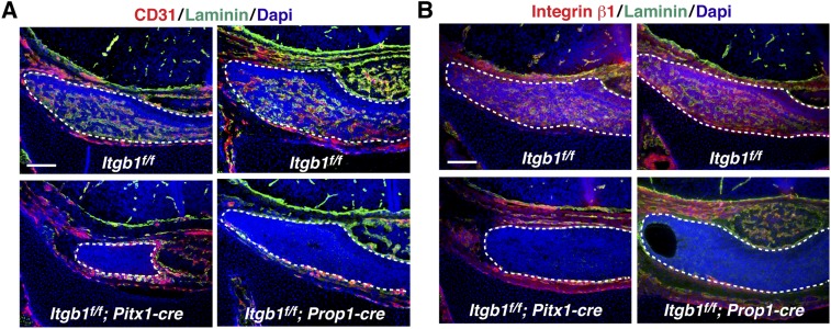
Fig. S4.
Absence of vasculature at p0 in both Itgb1f/f; Pitx1-cre and Itgb1f/f; Prop1-cre pituitary glands. (A) Laminin-rich vascular basement membranes have disappeared from pituitary glands in both Itgb1f/f; Pitx1-cre and Itgb1f/f; Prop1-cre knockouts by birth. (B) Integrin β1 expression is higher in vascular cells surrounded by laminin-rich vascular basement membrane than in surrounding epithelial cell parenchyma in controls. Coronal sections are shown in A and B. (Scale bars: 130 μm.)

