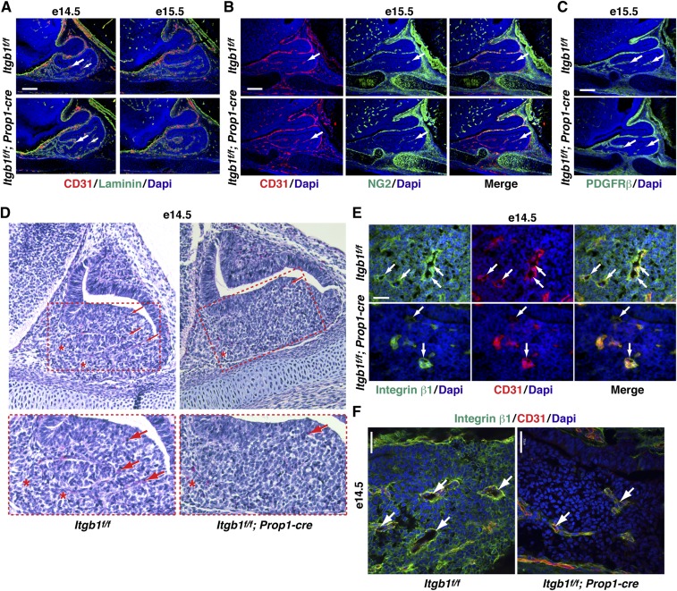
Fig. S6.
Reduced lumen formation in Itgb1f/f; Prop1-cre pituitaries at e14.5. (A) Endothelial cells are present (arrows) and surrounded by laminin-rich vascular basement membrane in e14.5 and e15.5 Itgb1f/f; Prop1-cre pituitary glands. (Scale bar: 130 μm.) (B) Pericytes immunostained with NG2 are associated with endothelial cells in e15.5 Itgb1f/f; Prop1-cre pituitaries. (Scale bar: 130 μm.) (C) Pericytes immunostained with PDGFRβ are associated with endothelial cells in midsagittal sections of e15.5 Itgb1f/f; Prop1-cre pituitaries. (Scale bars: 130 μm.) (D) H&E staining of parasagittal pituitary sections showed fewer RBCs (arrows) within the anterior lobe (red dashed lines) of Itgb1f/f; Prop1-cre pituitaries at e14.5. RBCs are present along the rostral aspect of both the Itgb1f/f and Itgb1f/f; Prop1-cre pituitary glands (asterisks). (Magnification: Top, 200×.) (E) Higher magnification images of lumens (arrows) from the section presented in Fig. 4A (Itgb1f/f, Top) and a section adjacent to the one shown in Fig. 4A (Itgb1f/f; Prop1-cre, Bottom). (Scale bar: 32.5 μm.) (F) Confocal images of lumens (arrows) in Itgb1f/f and Itgb1f/f; Prop1-cre pituitary glands at e14.5. (Scale bars: 30 μm.) Midsagittal sections are shown in A–C, parasagittal sections are shown in D, and coronal sections are shown in E and F.

