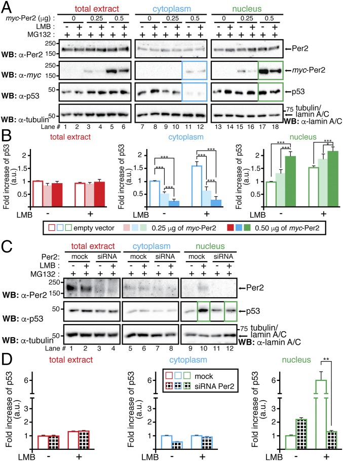
Fig. 5.
Per2 enhances p53 shuttling to the nucleus. (A) HCT116 cells were transfected with either myc-Per2 (0.25 or 0.5 μg) or empty vector (0) for 24 h before the addition of MG132 and/or leptomycin B (LMB). Lysates were subjected to subcellular fractionation, and total extract (Left), cytoplasmic (Middle), and nuclear (Right) fractions were analyzed for the presence of Per2, myc-Per2, and endogenous p53 by immunoblotting. Tubulin and lamin A/C were used as controls. (B) Endogenous p53 was quantified using Image Lab software/Gel Doc XR+ system and values normalized to tubulin or lamin A/C levels. (C) HCT116 cells were transfected with either scrambled siRNA (mock) or Per2 siRNA (siRNA) for 48 h before the addition of MG132 and/or LMB. Endogenous proteins were detected as indicated in A and quantified as in B. In B and D, data are represented as fold increase of p53 (in arbitrary units) compared with similar treatment in either vector-transfected or mock samples, respectively. Values are the mean ± SEM from three independent experiments. Statistical significance was determined by t test. *P ≤ 0.05; **P ≤ 0.005, ***P < 0.001.

