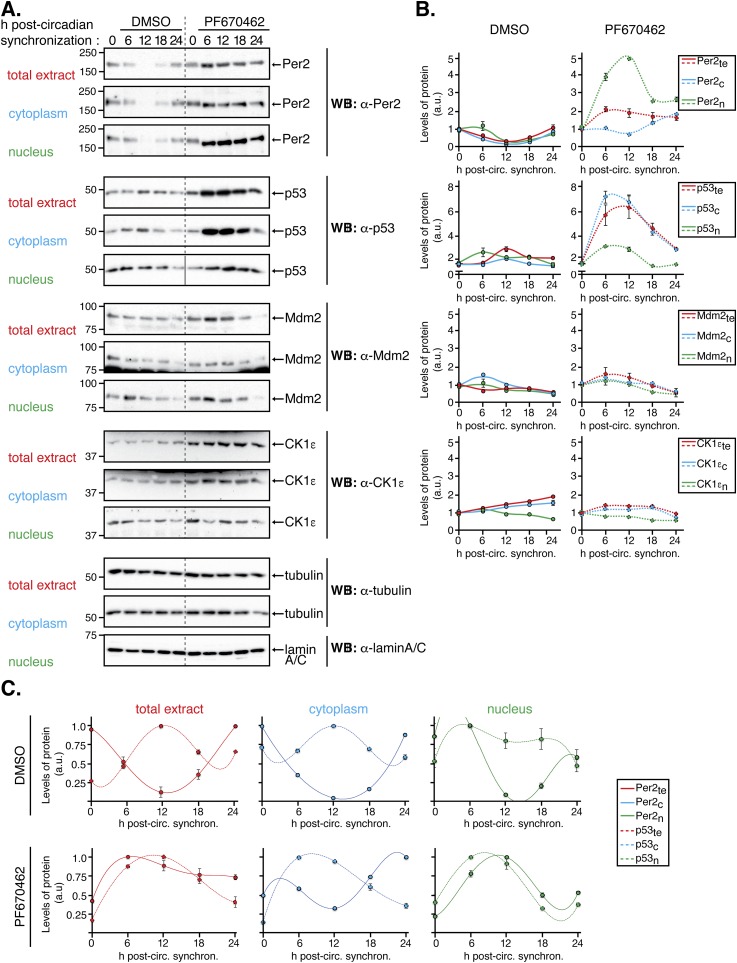
Fig. S2.
The Per2 and p53 phase relationship remains unaltered despite inhibition of CK1δ/ε. HCT116 cells were circadian synchronized by serum shock after which they were collected (t = 0) or maintained in serum-free media with DMSO (control, 0.2%) or PF670462 (1 µM) throughout the time course analyzed (t = 6–24 h). Cells were collected at the indicated times and lysates were subjected to subcellular fractionation as indicated in Materials and Methods. Levels of endogenous proteins were detected in total (1.2 × 10⁵ cells), cytoplasmic (0.63 × 10⁵ cells), and nuclear (3.15 × 10⁵ cells) extracts by immunoblotting using α-Per2, -p53, -Mdm2, and -CK1ε antibodies. Tubulin and lamin A/C were used as a loading and purity control for the cytoplasm and nuclear fractions, respectively. Immunoblot data originated from a single experiment that was repeated three times with similar results. (B) Protein bands from A were quantified using Image Lab software/Gel Doc XR+ system, and values were normalized to tubulin or lamin A/C levels (loading controls), and data [in arbitrary units (a.u.)] were represented as the mean ± SEM from three independent experiments. Plots were grouped by the protein’s class and expression in its various compartment in cells treated with either DMSO (Left) or PF670462 (Right) treatment. (C) Data from B was replotted to better show the conservation of the phase relationship between Per2 and p53 in the indicated compartments.

