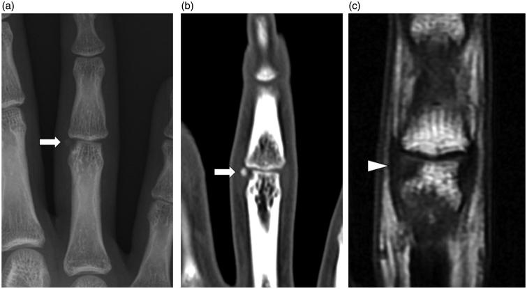
Figure 1.
Plain x-ray, computed tomography (CT), and magnetic resonance imaging (MRI) of the right fourth finger. (a) Plain X-ray, (b) coronal CT section, (c) T1-weighted MRI image. Plain X-ray and CT images show a bone defect on the radial side of the head of the proximal phalanx and a fragment (white arrows). MRI image shows an osteochondral lesion without associated radial collateral ligament tear (white triangle).

