
Fig 6.
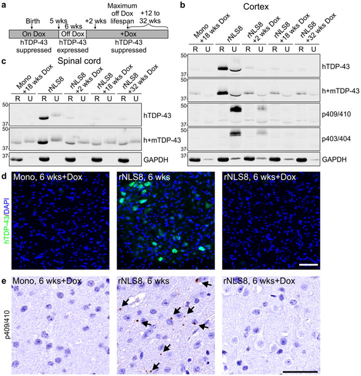
Soluble and insoluble TDP-43 are rapidly cleared from brain and spinal cord of rNLS8 mice upon re-introduction of Dox. a Schematic of experimental design. b, c Levels of hTDP-43 and h+mTDP-43 in RIPA-soluble (R) and RIPA-insoluble/urea-soluble (U) protein fractions of rNLS8 mouse cortex and spinal cord at 6 weeks and at subsequent time points back on Dox (+Dox), with tetO-hTDP-43-ΔNLS monogenic (Mono) littermate control. p403/404-TDP-43 and p409/410-TDP-43 were rapidly eliminated from the U fraction. Approximate molecular weight markers in kDa are shown on the left and GAPDH is the loading control. d hTDP-43 was eliminated from spinal cord MNs in rNLS8 mice +Dox, and e p409/410-TDP-43 inclusions were detected in the motor cortex in rNLS8 mice at 6 weeks, but were not detected in rNLS8 mice +Dox (images of 6 weeks off Dox +18 weeks back on Dox are shown). Scale bars d 100 μm, e 50 μm