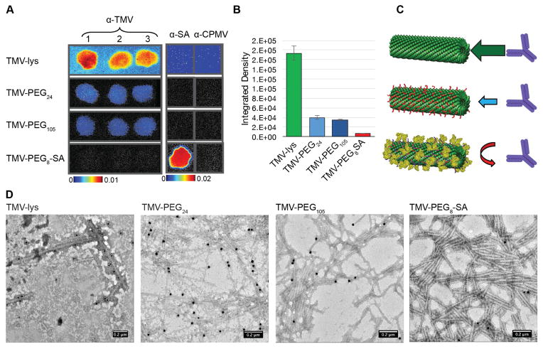
Figure 2. Immune recognition of TMV-PEG8-SA.
A, Immune recognition of fluorescent ‘naked’ and ‘stealth’ TMV particles by α-TMV and α-SA antibodies using dot blots. The binding of particles to α-TMV antibody spotted on the membrane is decreased with PEG coatings and was effectively prevented using SA coatings. B, Quantitative densitometric analysis of the dot-blots (A). C, Schematic representation of the antibody-recognition of the various TMV-based particles. D, TEM images of TMV formulations after immunogold staining using α-TMV and gold-labeled secondary antibodies. HSA was used to prepare the TMV-PEG8-SA particles.

