FIG. 3.
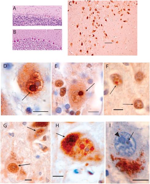
SCA12 histopathology and intranuclear neuronal inclusions. (A) SCA12 cerebellar cortex (H & E staining) showing Purkinje cell loss. (B) Cerebellar cortex, normal control: H & E staining. Scale bar = 200 μm. (C) SNpc, immunostained for ubiquitin, showing melanin-containing neurons that exhibit normal density, size, and appearance for a 66-year-old subject. Scale bar = 100 μm. (D) Purkinje cell with a ubiquitin positive, round, intranuclear inclusion (arrow) adjacent to a similarly sized pale blue nucleolus. (E) A different Purkinje cell containing a ubiquitin-positive, round, intranuclear inclusion (arrow). (F) Two neurons (arrows) in motor cortex with multiple, ubiquitin-positive, intranuclear inclusions distinct from the nucleolus (stained blue). (G) Two neurons in the SNpc. The upper neuron contains two ubiquitin-positive, intranuclear inclusions adjacent to each other and separate from the nucleolus. The lower neuron contains one ubiquitin-positive intranuclear inclusion adjacent to the nucleolus. (H) SNpc neuron with at least four round, ubiquitin-positive, intranuclear inclusions surrounding the nucleolus. Arrow points to cytoplasmic melanin. (I) SNpc neuron with cytoplasmic melanin inferiorly. Pale blue nucleolus indicated with arrowhead. Arrow points to a smaller, round inclusion that is negative for 1C2 immunostaining (expanded polyglutamine repeats) and is faintly eosinophilic. Sections D to I are immunostained for ubiquitin and counterstained with H & E. Section I is immunostained with 1C2 antibody and counterstained with H & E. Scale bars are 10 μm in sections D to I.

