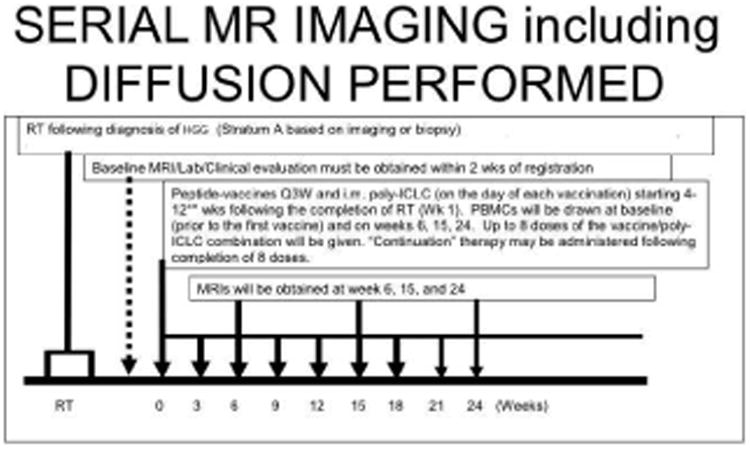
Figure 1. Example of Timing of MRI scans for New Diagnosis of High-Grade Pediatric Glioma treated with Radiation and Serial Peptide Based Vaccine Therapy.

The time of conventional MRI during the course of peptide-vaccine therapy for this particular strata (A) of the vaccine study was approximately every 6 weeks after initiation of therapy. Strata A of included new diagnosis of high-grade gliomas based on imaging (DIPG) or biopsy and included initial radiotherapy followed by peptide-based vaccine. Note, additional time points of imaging were obtained during clinical pseudoprogression. The timing of serial MRI was different for different strata. Diffusion imaging was integrated with all conventional MRI scans. MRS spectroscopy and perfusion MR were performed in conjunction with only certain conventional MRI scan for logistical reasons.
