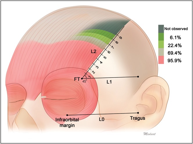
Figure 3.

Distance from FT to the musculoaponeurotic junction of the lateral border of the frontalis with the galea aponeurotica. Shading indicates the different running patterns of the lateral border of the frontalis muscle. The lateral border of the frontalis extended at least to section 5 in 49 of 49 cases (100%), to section 6 in 47 cases (95.9%), to section 7 in 34 cases (69.4%), to section 8 in 11 cases (22.4%), and to section 9 in 3 cases (6.1%).
