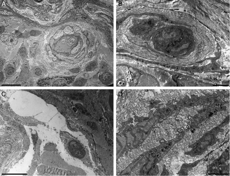
Figure 5.
Transmission electron microscopy of the skin after treatment with fat plus PRP. (A) Perivascular infiltrates. (B) Reduplicated vascular basement lamina. (C) Oedema among the connective fibers of the dermis. (D) Activated vascular endothelium is visible. Scale bars: (A) 10 µm, (B) 2 µm, (C) 5 µm, (D) 2 µm.

