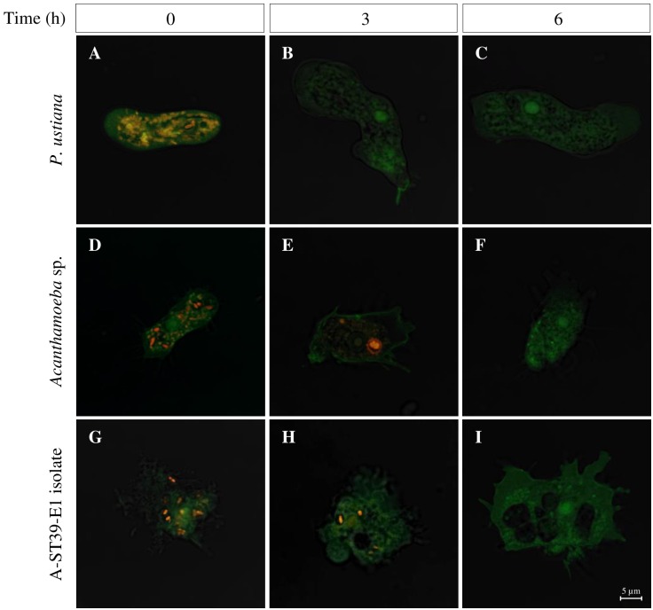
Fig 3. B. pseudomallei is internalized into amoebae but could not resist digestion.
CLSM micrographs show the internalized B. pseudomallei in P. ustiana (A-C), Acanthamoeba sp. (D-F) and isolate A-ST39-E1 (G-I) at 0, 3 and 6 h after kanamycin treatment. Orange fluorescence represents CellTracker™ Orange-B. pseudomallei and green fluorescence indicates the amoebae stained with FITC-ConA for visualization.

