. 2016 Nov 17;5:e17896. doi: 10.7554/eLife.17896

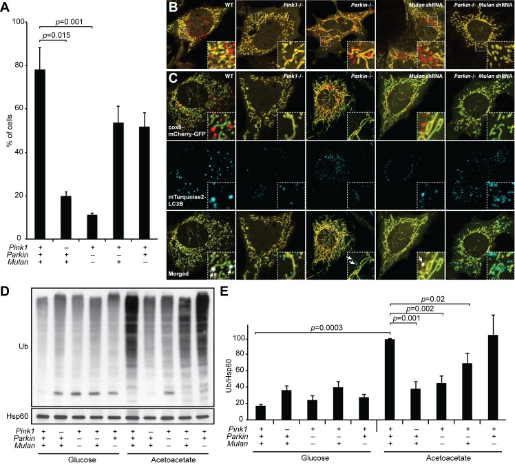
Figure 4. MUL1 and PARKIN have redundant functions in OXPHOS-induced mitophagy.

(A) Quantification of red-only puncta in cells grown in acetoacetate-containing medium. Presence (+) or absence (-) of Pink1, Parkin, or Mul1 is indicated. Error bars indicate SD of three biological replicates, p=0.015 (Pink1), p=0.0011 (Parkin-/- Mulan shRNA) (Student’s t-test). (B) Mitophagy in wild-type and mutant cells. Cells stably expressing Cox8-EGFP-mCherry were grown in acetoacetate-containing medium and imaged by fluorescence microscopy. (C) Co-localization of LC3B with mitophagy intermediates. Wild-type and mutant cells were retrovirally transduced with mTurquoise2-LC3B, grown in acetoacetate-containing medium and imaged by fluorescence microscopy. Examples of LC3B co-localization with mitophagy intermediates are indicated by arrows. (D) Accumulation of polyubiquitinated proteins in mitochondria. Cells were grown in the indicated medium, and mitochondria were isolated by differential centrifugation. Mitochondrial lysates were analyzed by Western blot for pan-Ubiquitin. HSP60 is a loading control. (E) Quantification of polyubiquitinated proteins in mitochondria. Three independent experiments were quantified by densitometry and averages are shown. Ubiquitin level was normalized to HSP60. Error bars indicate SD, p=0.0003 (WT Glu vs. Ac), p=0.0011 (Pink1-/-), p=0.0016 (Parkin-/- Mulan shRNA), p=0.0206 (Parkin-/-) (Student’s t-test).

DOI: http://dx.doi.org/10.7554/eLife.17896.008

Figure 4.

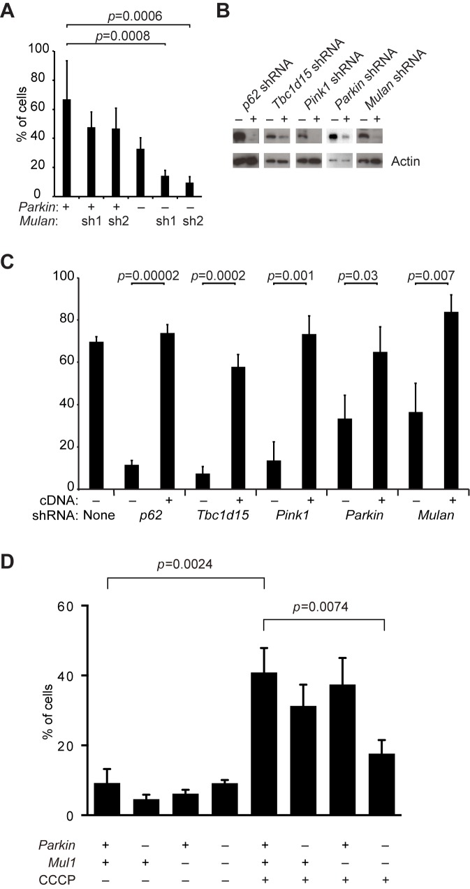
Figure 4—figure supplement 1. Defective mitophagy in Parkin/Mul1-deficient cells.

Figure 4—figure supplement 1.

(A) Requirement for Parkin/Mul1 in mitophagy. Quantification of red-only puncta in cells grown in medium containing acetoacetate. Wild-type (+) or Parkin knockout (-) cells were transduced with one of two independent Mulan shRNAs as indicated. Error bars indicate SD from two biological replicates. p values are from the Student’s t-test. (B) Western blot analysis of shRNA knockdowns. Cells were transduced with the indicated shRNA (+) or not (-) and blotted with corresponding antibody. For analysis of the Parkin shRNA, MEFs overexpressing EGFP-Parkin were used, because endogenous Parkin is below the detection limit of the antibody. Actin was used as a loading control. (C) Rescue of mitophagy with shRNA-resistant cDNA expression. Cells expressing the indicated shRNA were transduced with shRNA-resistant cDNA constructs as indicated (+). The cells were grown in acetoacetate containing medium, and mitophagy was quantified as described in Figure 4. Error bars indicate SD from three biological replicates. p values are from the Student’s t-test. (D) Dependence of CCCP-induced mitophagy on Parkin and Mul1. MEFs of the indicated genotypes were incubated with (+) or without (-) CCCP (10 μM) for 6 hr, and mitophagy was quantified. Error bars indicate SD from three independent experiments. p values are from the Student's t-test.